
Study skins on their way into the collection (Image: Mareike Vennen/MfN. All rights reserved.)
Content Warning: The following text and video contain explicit pictures of preparing a dead bird’s body that may be disturbing to some viewers.
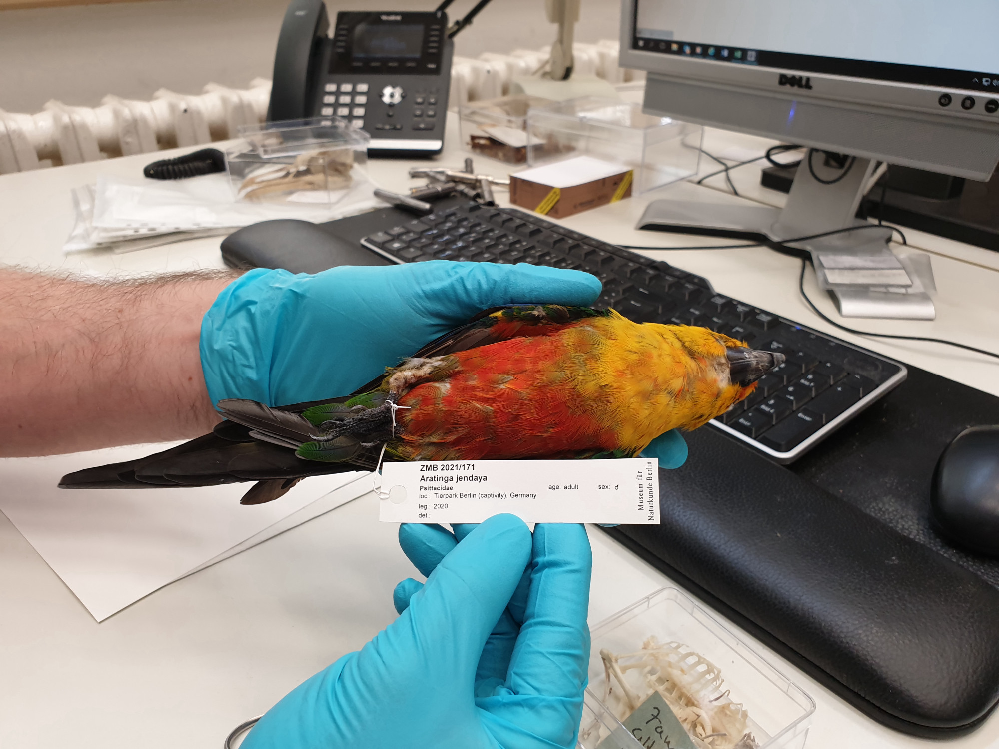
In autumn 2020 and spring 2021, our team observed and documented the preparation of a bird study skin at the Museum für Naturkunde Berlin. The bird in question was a male Jendaya parakeet (Aratinga jandaya) that had hatched on 13 June 2006 at Berlin Tierpark and had been sent from the Tierpark to the Museum für Naturkunde Berlin after its death on 29 August 2020.1
This video illustrates the work of Christin Scheinpflug, a preparator at the Museum für Naturkunde Berlin. It shows her turning a Jendaya parakeet from the Tierpark Berlin into a study skin and a partial skeleton for the museum’s Bird Collection. Preserving the remains and mounting them for the research collections of the museum is a long process that involves many steps and many transformations. In documenting these steps the video provides insights into how animals can turn into scientific objects of study. (Video: Filippo Bertoni/MfN. All rights reserved.)
As our project investigates how zoo animals become museum collection objects, we were keen to record the individual moments of this transformation. To this end, we visited specimen preparator Christin Scheinpflug in her workshop over several months.2 Once the preparation was done, we were also able to follow the collection manager Pascal Eckhoff as he entered information about the study skin into the database and catalogued it as part of the Bird Collection.
Whether Tierpark and Zoo animals are actually sent to the Museum für Naturkunde Berlin depends on a number of questions, like what kind of animals they are, which species they represent, whether the collection already has specimens of that species in good conditions, and how rare the species is. How well-preserved an  animal’s body is can also be crucial for deciding whether it will be prepared as a specimen. This also determines how it will be stored at the museum – whether as a skeleton or a study skin (with a partial skeleton) for the scientific collection, or as a taxidermy piece in the exhibition.
In this case, it was decided to prepare a study skin and a partial skeleton.3 Because a study skin is a scientific object, it is important to utilise and preserve the original body parts. For this purpose, a study skin was made from the skin of the Jendaya parakeet together with its feathers, beak, legs, and feet (including its lower legs and foot bones), as well as its wings (including its falanx, forearm, and shoulder bones). A partial skeleton refers to bone structures that do not add up to a complete skeleton because parts are missing or have been used, for example, in making the study skins. In the case of our Jendaya parakeet, a partial skeleton was crafted using the cervical spine and the trunk skeleton left from the earlier preparation.4 All other parts of the skeleton – the femurs, skull, falanxes, arms, and shoulders – remained in the study skin.
In ornithology, the process of crafting a study skin and making a partial skeleton are considered part of a single practice. Because important parts of the skeleton, such as the skull and fibula, are used for the skin, the curator must decide prior to preparation whether to make a complete skeleton or a study skin with a partial skeleton. In our case, the bird’s ribs had already been opened up to extract the organs during the first dissection. Also, parts of the chest muscles had been removed.
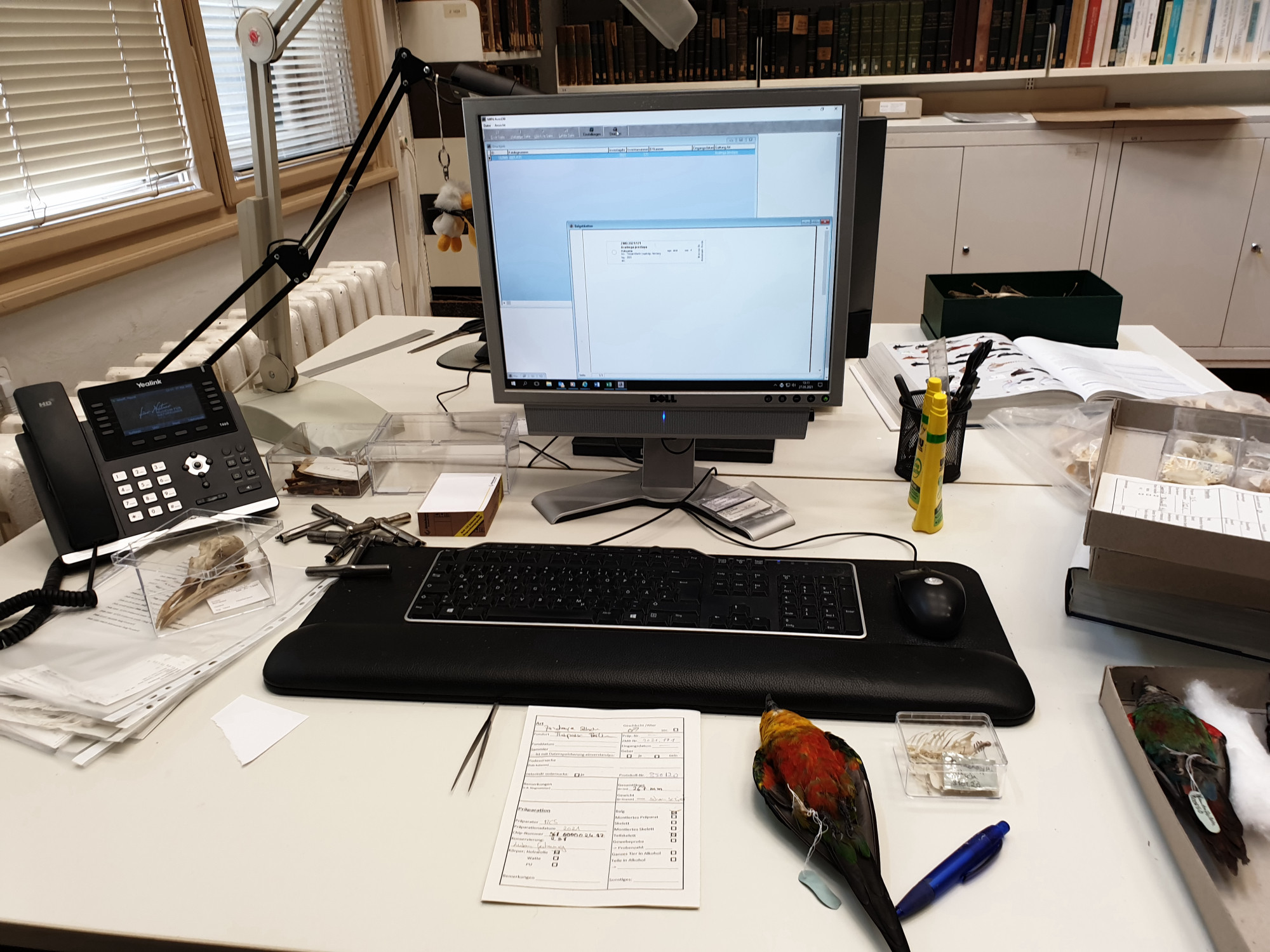
After dissection, the bird was immediately deep frozen and tagged with the letters ‘MFN’ (Museum für Naturkunde Berlin). After its arrival at the museum and before its preparation, the bird was stored in the freezer of the museum’s preparation workshop. The first step in the bird’s scientific preparation took place in the museum’s dissection room, where the animal’s body was first thawed in a solution of salt and Supralan. After that,  scientific data was collected, including data about the animal’s
  dimensions, accession number, date of arrival, and chip number.5 The preparator entered this information into a
  data sheet.
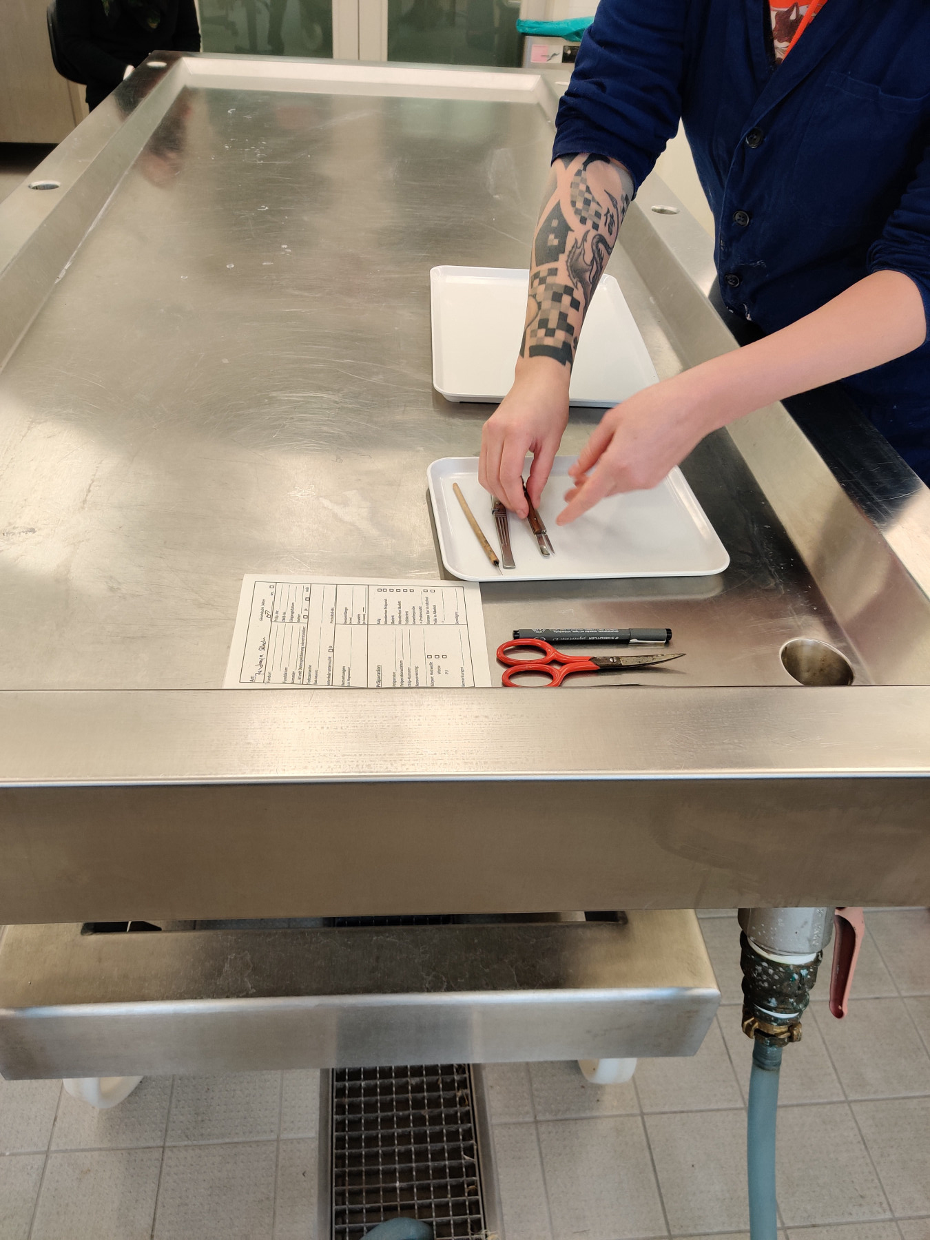
Data sheet and instruments for measuring the specimen, obtaining the skin, and preparing the partial skeleton (Image: Filippo Bertoni/MfN. All rights reserved.)
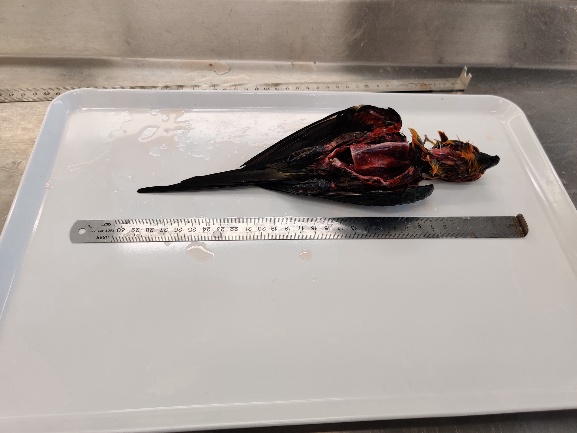
Measuring the bird’s body (Image: Filippo Bertoni/MfN. All rights reserved.)
A  data sheet is prepared for each animal. This includes information about materials and techniques as well as some key information about the bird and the date of preparation.
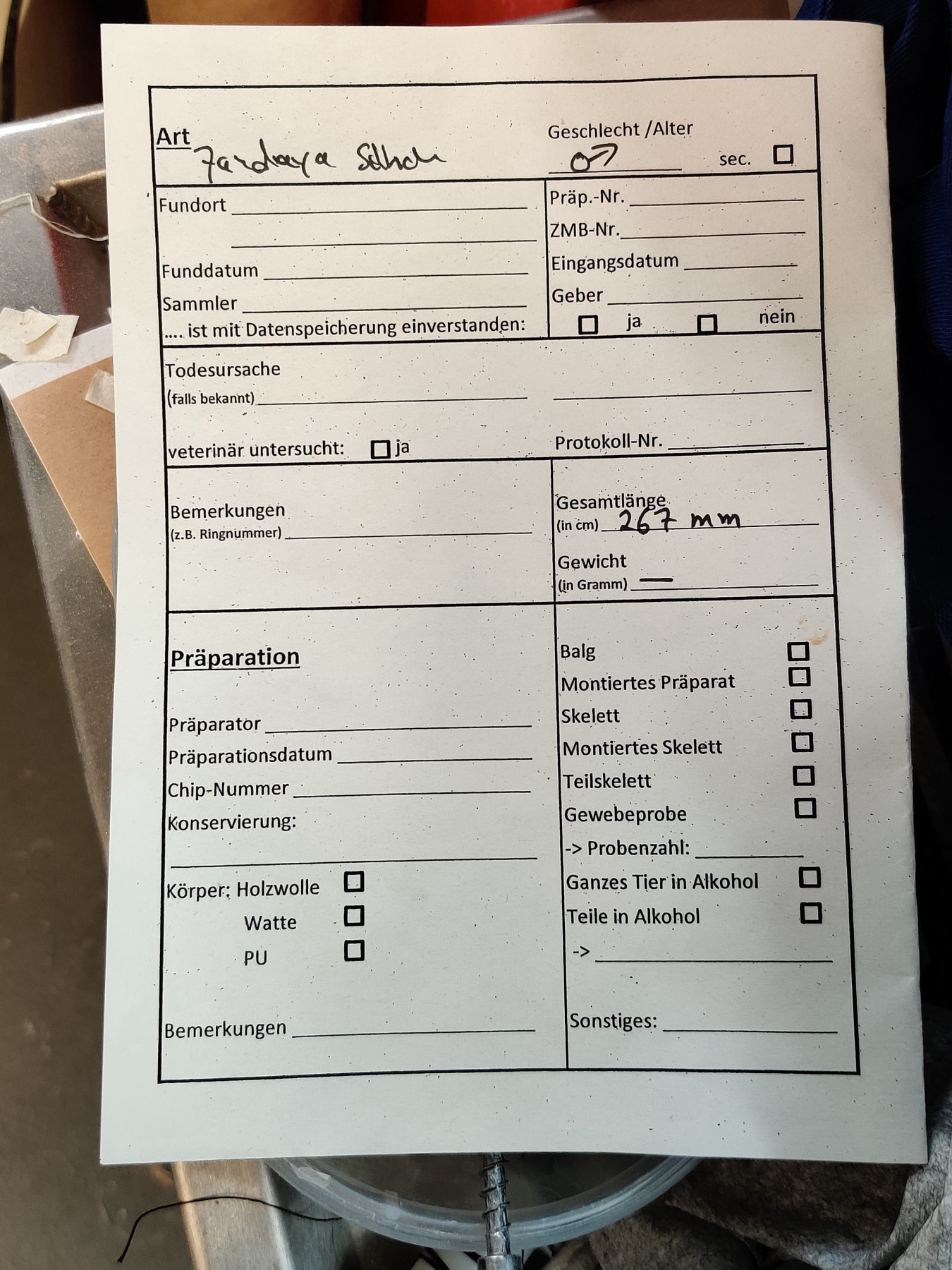
Data sheet; the information from the data sheet will later be entered in the collection catalogue. (Image: Filippo Bertoni/MfN. All rights reserved.)
In this case, the live bird had already been chipped for identification purposes while still at the Tierpark. This means that a transponder with a chip number on it (968000002487298) had been implanted at about the level of the trachea.6 This number links the bird to the ![]() ZIMS (Zoological Information Management Software) database, which many zoos use to coordinate their breeding and zoo-keeping practices. This digital repository is where you can find further information about the individual animal if the zoo has provided it. The transponder was removed during the preparation process and stored together with the data sheet before being placed back into the prepared skin. This is done because by virtue of containing information about the (zoo) animal, the tag is considered part of it. It thus became part of the study skin and part of the scientific collection.
ZIMS (Zoological Information Management Software) database, which many zoos use to coordinate their breeding and zoo-keeping practices. This digital repository is where you can find further information about the individual animal if the zoo has provided it. The transponder was removed during the preparation process and stored together with the data sheet before being placed back into the prepared skin. This is done because by virtue of containing information about the (zoo) animal, the tag is considered part of it. It thus became part of the study skin and part of the scientific collection.
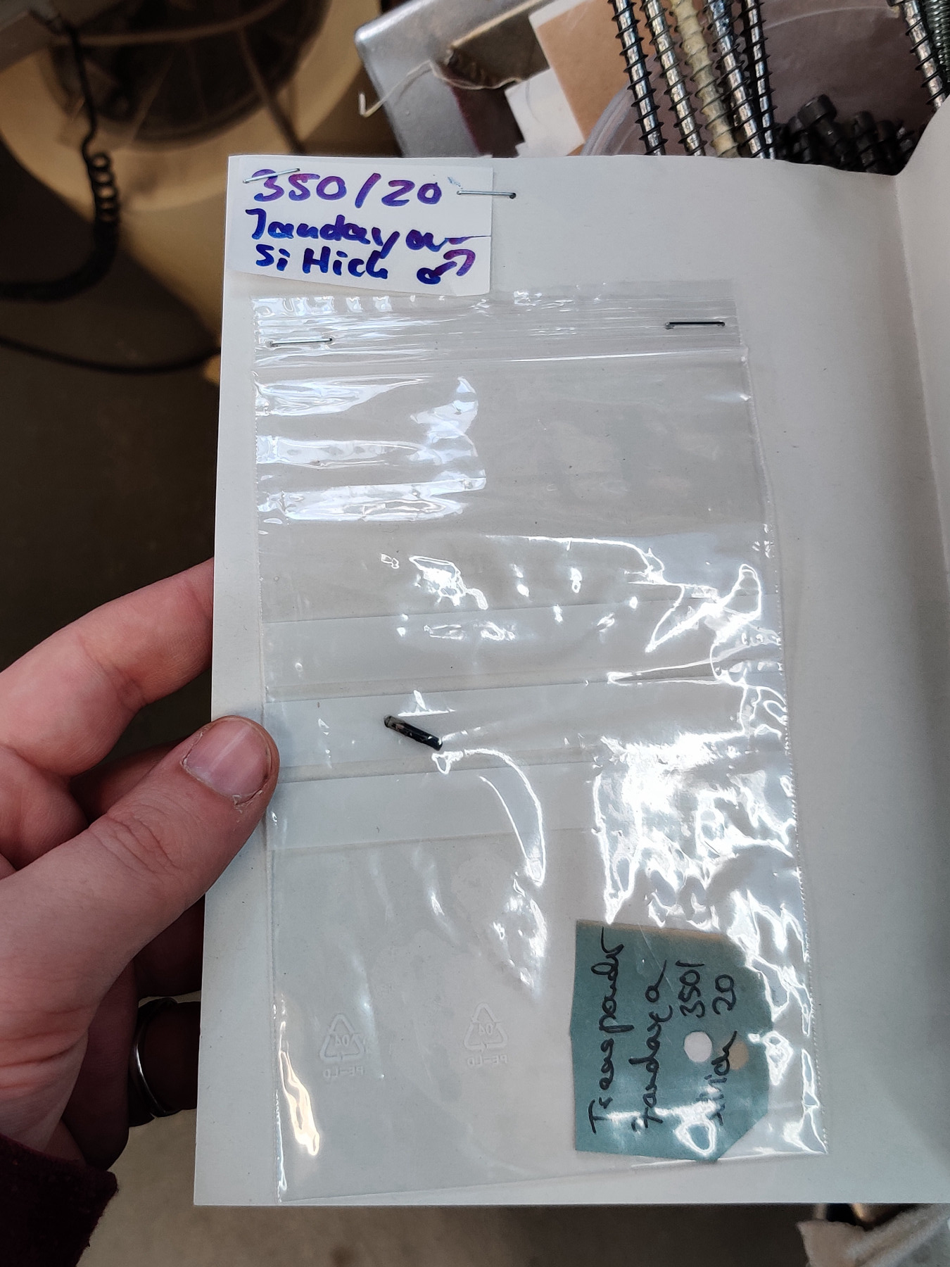
This is how the transponder is stored until it is placed back into the study skin at the end of the preparation. (Image: Filippo Bertoni/MfN. All rights reserved.)
In order to make the study skin, the preparator first has to remove the skin using a scalpel and a pair of scissors in order to separate it from the flesh tissue. Afterwards, the skin is mechanically cleaned of fascia and fat remains. The muscles and tendons are then removed from the remaining bone structures (fibulas, skull, shoulder and forearm) – this process is also referred to as cleaning or ‘defleshing’. Removing the fat and fascia from the skin and defleshing the bones is an important part of the preparator’s work, as the smell of fat and tissue remains attract insects, which can potentially  damage the study skin in the collection.
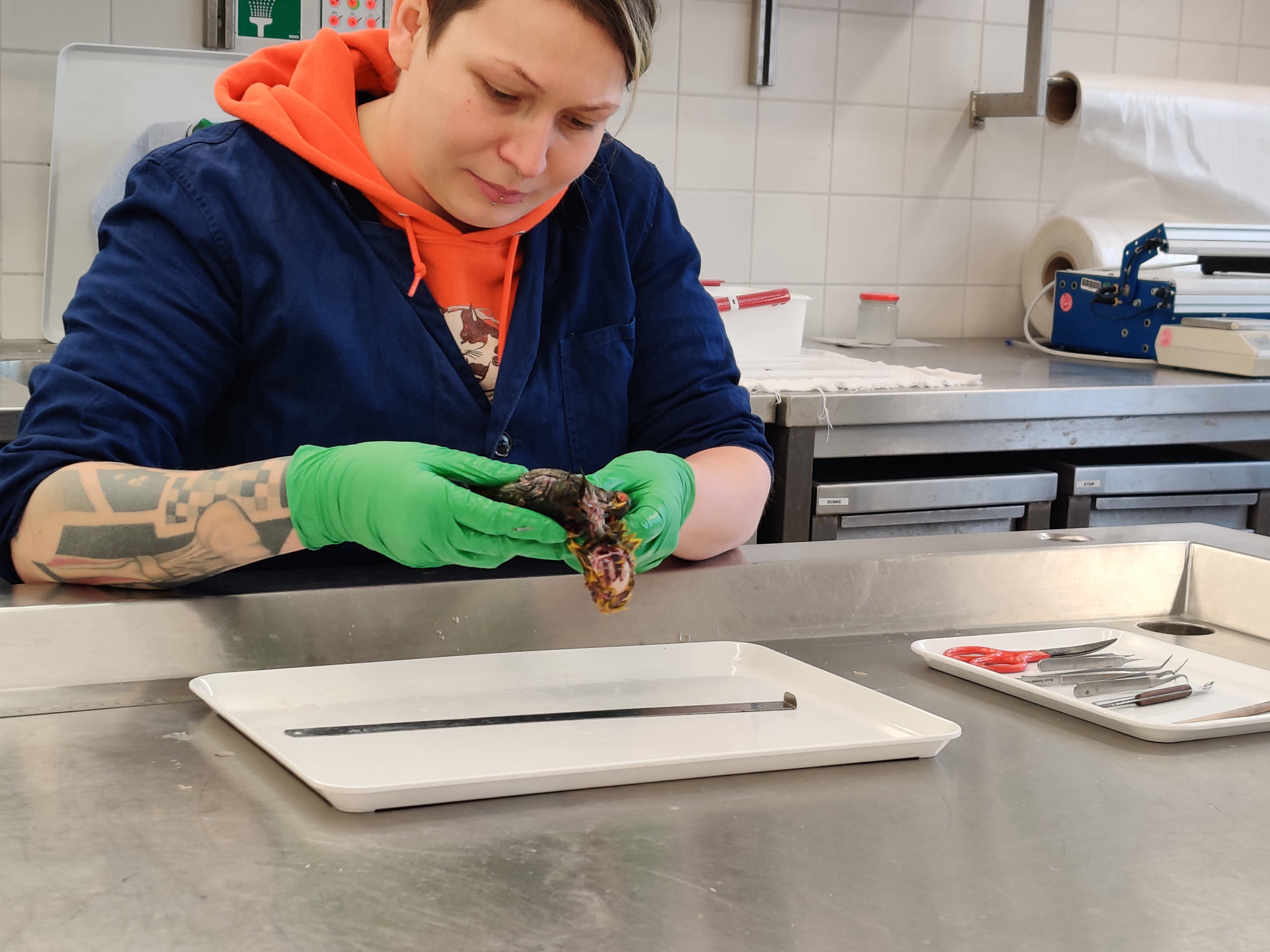
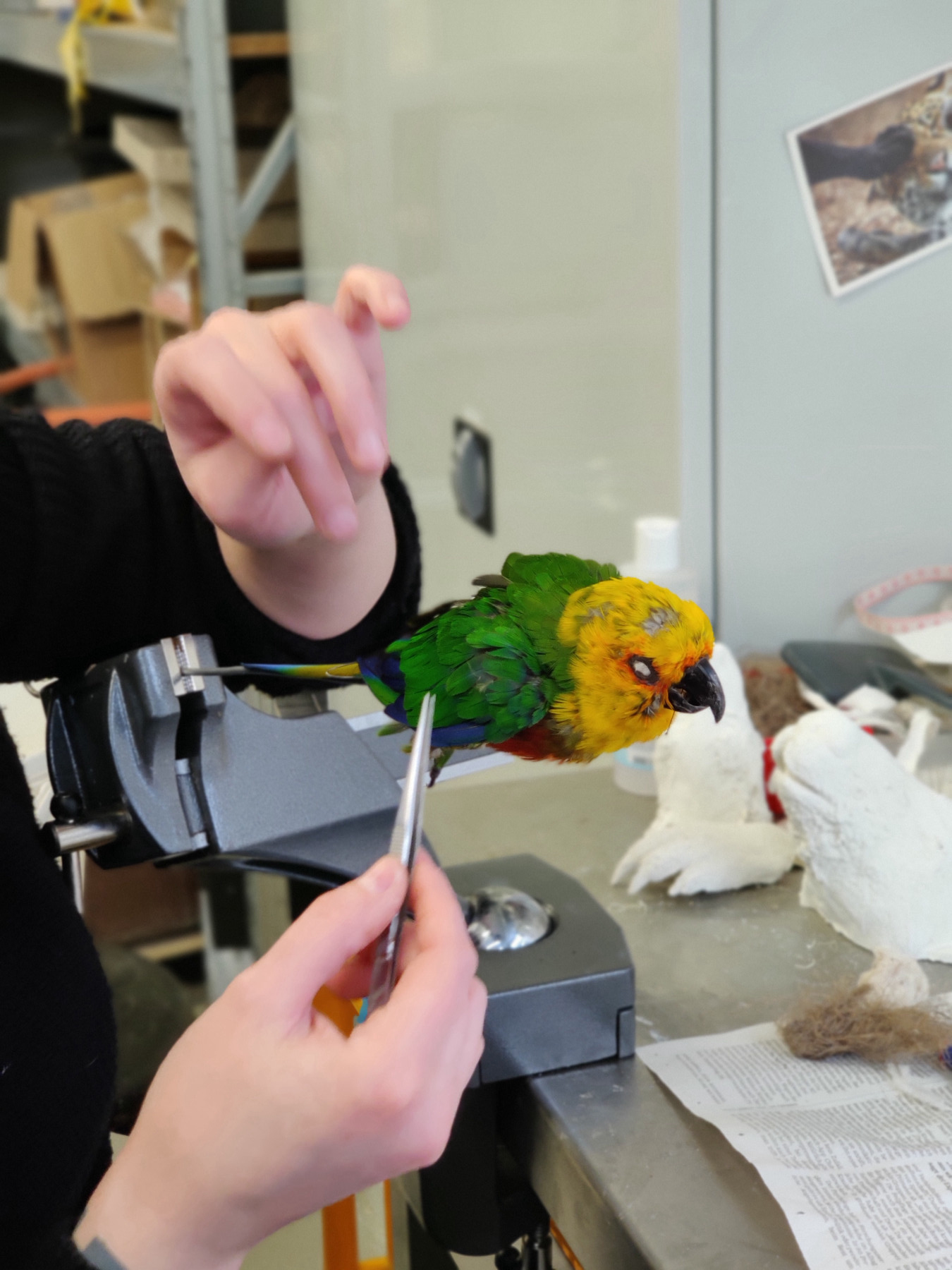
The museum preparator Christin Scheinpflug examines the bird’s body. (Image: Filippo Bertoni/MfN. All rights reserved.)
While the remaining tissue is removed and later professionally  disposed through incineration, the bird skin is washed multiple times in water, Supralan, and shampoo, and then once again stored in a plastic bag in a freezer until tanning.7
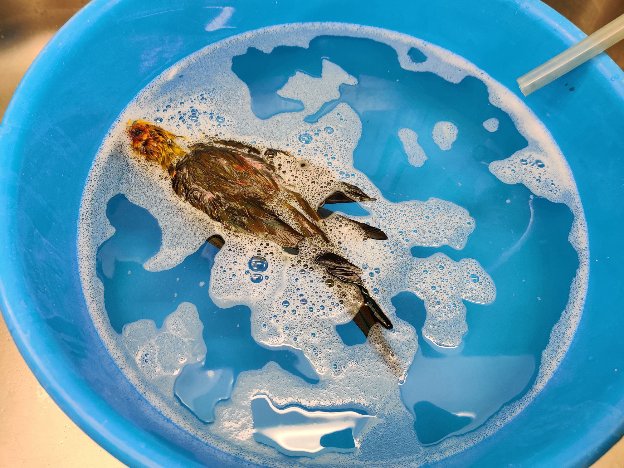
A water bath with Supralan and salt to clean the skin and any potentially dirty feathers (Image: Filippo Bertoni/MfN. All rights reserved.)
During the tanning process, where tanning agents are used for long-term preservation of animal skins, the body is first softened in a salt and Supralan solution. The tanning process takes place in several stages and fixes the tanning agent in the skin. This takes about one day. The damp skin is stored on a rack in the fridge overnight so that tanning can be finished the next day in a neutralisation process. Preparators always keep a log of the tanning process.
During the tanning process, the pores are softened in a lye solution and a tanning agent is then fixed in the skin. (Image: Filippo Bertoni/MfN. All rights reserved.)
While the bird’s skin was tanning, Christin Scheinpflug prepared the artificial body to which the study skin would later be attached, crafting it out of wood wool, a natural material. She compressed and wrapped the wood wool tightly using cotton thread, allowing a solid shape to take form. She would later be able to pull the tanned skin over this body.
The artificial body for the study skin is made from wood wool and wire and measured against the dimensions of the bird’s body. (Image: Filippo Bertoni/MfN. All rights reserved.)
The preparator marked the places on the wood-wool body where the wings and lower legs would later be attached. Using a stronger cotton thread, she sewed the wood-wool body up at one end and fixed the neck to it, which was made of wrapped cotton wool instead of wood wool (for more flexibility).
For the final steps – assembling the bird, sewing it up, and putting the feathers in place – we moved from the dissection room to Christin Scheinpflug’s workplace in the museum’s preparation workshop. The assembly process always begins with the wiring of the wing bones and fibulas. A wire is wrapped tightly around the bones using a cotton thread, so that the bones can later be anchored in the wood-wool body. Afterwards, the preparator pulled the tanned skin over the artificial body and connected the bird’s neck and skull using a wire. She also applied Dextrinton (a mixture of Dextrin and sawdust) in some places to stabilise the connection between the skull and the neck. Afterwards, the preparator inserted and pinned the wing bones including the wire into the artificial body, then did the same to the lower legs. She subsequently sewed the skin to the wood wool body.8 As she sewed the body up, she also returned the transponder back into the chest cavity of the wood-wool body.
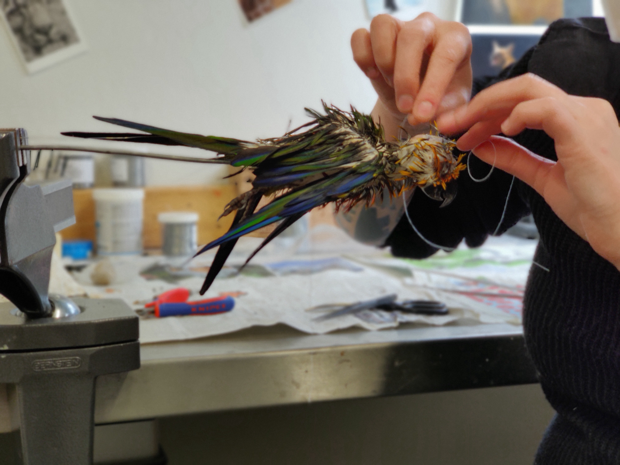
After tanning and inserting the wood wool body, the preparator sews up the skin. (Image: Filippo Bertoni/MfN. All rights reserved.)
A metal pole inserted into the bottom of the wood-wool body serves as a handle for holding the study skin in place during the preparation process. In particular, it makes sewing easier and protects the feathers.
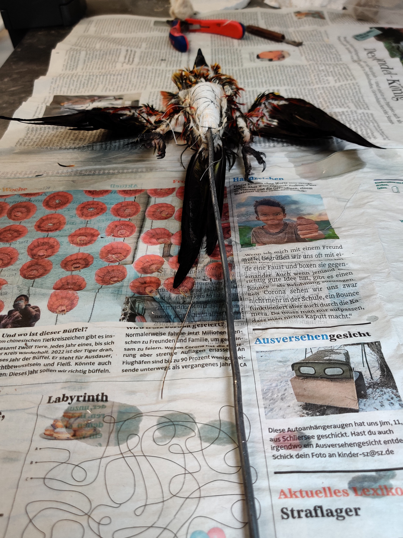
Wet study skin with wood wool body and metal handle inserted (Image: Filippo Bertoni/MfN. All rights reserved.)
After the feathers have been blow-dried, they have to be put arranged, which takes several hours. This means aligning the feathers so that they appear in the same way they would on a living bird.
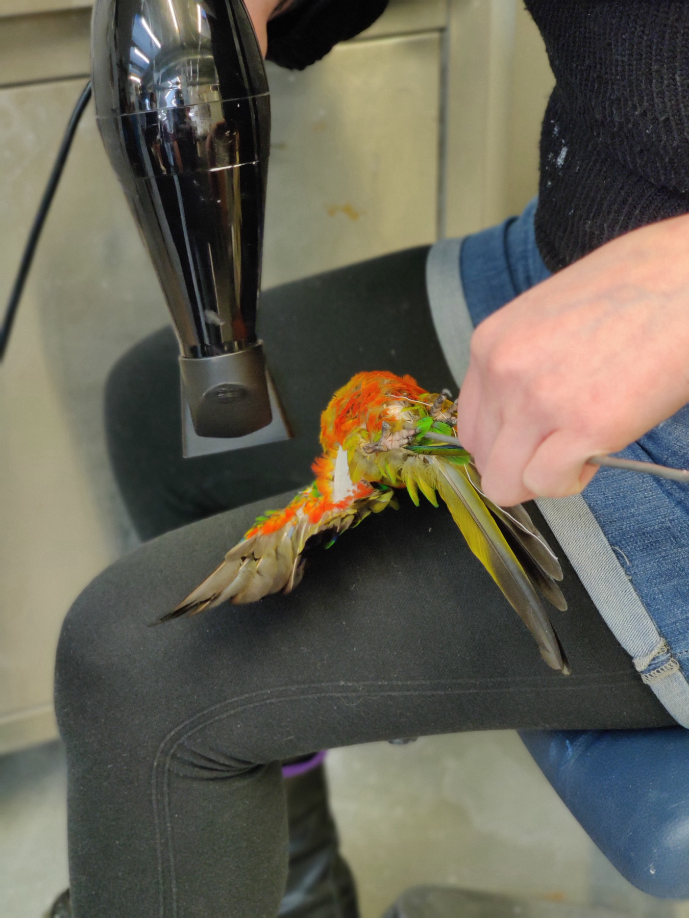
Blow-drying and arranging the feathers (Image: Filippo Bertoni/MfN. All rights reserved.)
In order to produce a partial skeleton, the original bird body now has to be completely ‘defleshed’. But unlike the skull bones, it will not be placed back inside the study skin. The defleshed bird body was then placed in a salt solution to ‘bleed’ it. Due to the size of the bird’s body, the preparator decided to clean the bones by enlisting the help of dermestid beetles. This meant placing the various parts of the skeleton into a container filled with dermestid beetles in a heated cabinet for several days.9 These scavenger beetles do not leave any tissue on the bones.
Producing a partial skeleton inside a warming cabinet with the help of dermestid beetles (Image: Filippo Bertoni/MfN. All rights reserved.)
Afterwards, the skeleton was frozen once more (in order to kill any remaining beetles on or inside the bones), defatted, and bleached. It was then ready for the collection. We accompanied Christin Scheinpflug with the study skin, the partial skeleton, and the data sheet from the museum’s preparation workshops to the Bird Collection.
Study skins on their way into the collection (Image: Mareike Vennen/MfN. All rights reserved.)
There, collection manager Pascal Eckhoff entered the information from the data sheet into the digital collection database.10 We noticed that this animal was not the first specimen of Aratinga jandaya in the museum’s Bird Collection. In 1895 and 1923, the collection had received two other specimens for its study skin collection. Moreover, the ornithological collection also contains other Jendaya parakeets  preserved in alcohol as well as a skeleton.
In the Bird Collection, the study skin is entered into the database. (Image: Mareike Vennen/MfN. All rights reserved.)
Label to identify the study skin (Image: Mareike Vennen/MfN. All rights reserved.)
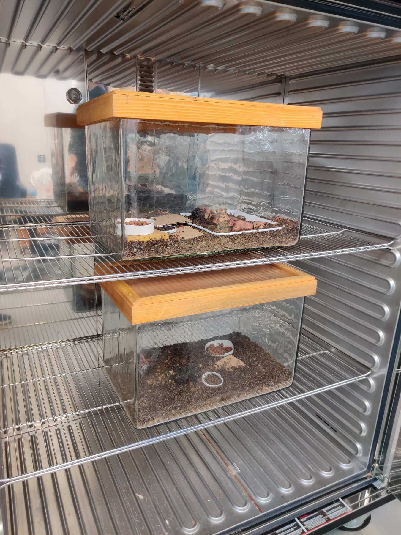
In the end, the bird was entered into the collection database under the inventory number ‘ZMB 2021.171’, while the study skin and the partial skeleton were put away in a drawer in the scientific collection. They have thus officially joined the collection of the Museum für Naturkunde Berlin.
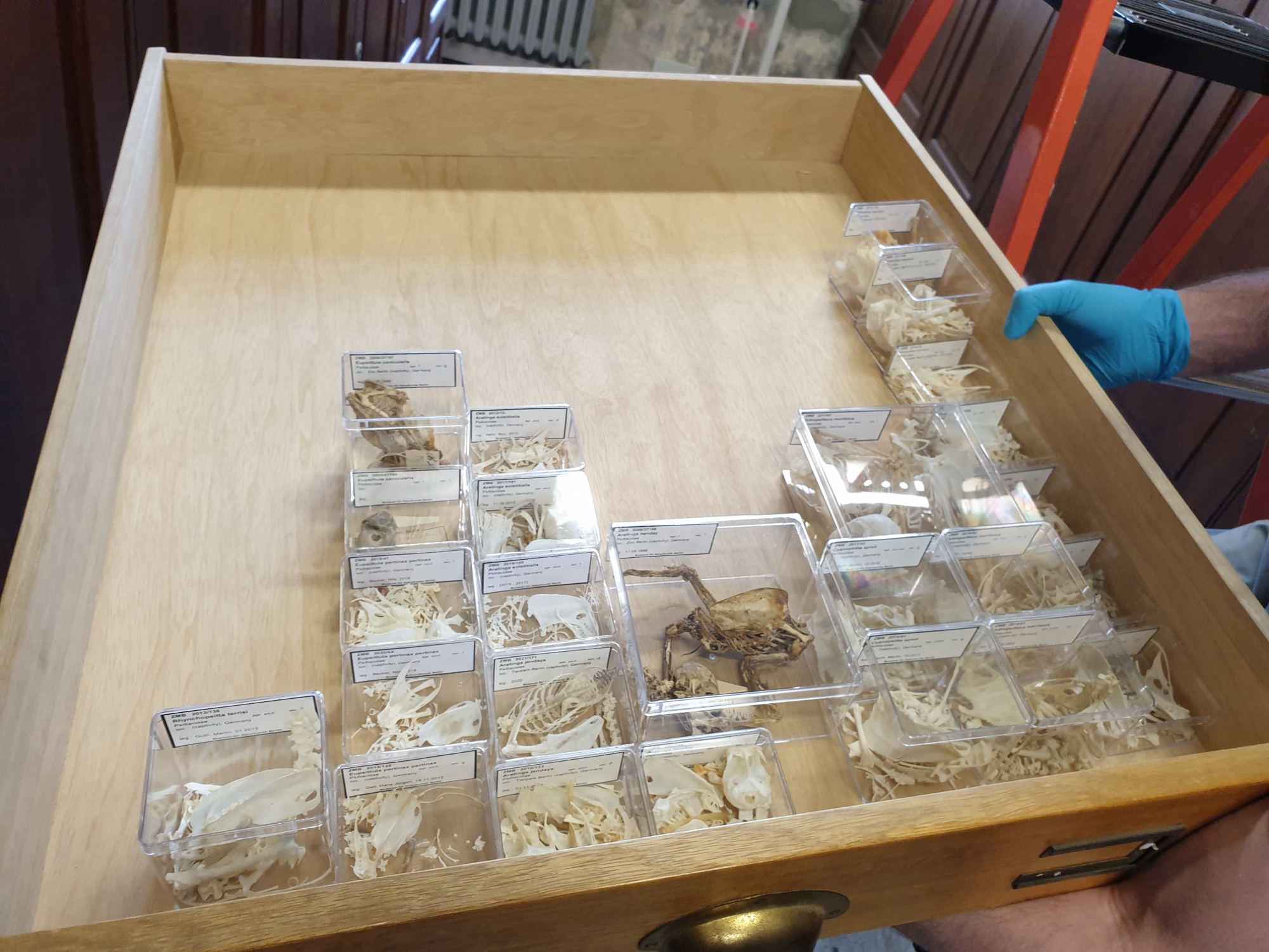
Storing and classifying the partial skeleton in the collection (Image: Mareike Vennen/MfN. All rights reserved.)
- Data like the date of death, age, and the date it hatched are collected in the ZIMS database at the Tierpark. According to the database, the parent animals are unknown. The group of Jendaya parakeets at the Tierpark currently (in October 2021) comprises eleven individuals that breed regularly. The avian subject of this article was the oldest in the group. Jendaya parakeets generally reach the age of 20 in zoos.↩
- The various preparation steps often do not take place all at once, but with interruptions, meaning that the process can stretch out over several weeks or months. We would like to thank Christin Scheinpflug for letting us take part in the process, for answering our numerous questions, and for providing us with all information needed.↩
- Unlike a taxidermy, a study skin has the feathers, beak, legs, and feet attached to it and is pulled over an artificial replica of the animal’s body. It can be stored in a drawer. In today’s scientific collections, study skins are the main type of specimen used. On working in and with the Bird Collection at the Museum für Naturkunde Berlin, see Sylke Frahnert, M. Päckert, D. T. Tietze, and T. Töpfer. “Aktuelle Schwerpunkte sammlungsbezogener Forschung in der Ornithologie”. Vogelwarte 51, no. 3 (2013): 185-191.↩
- The torso skeleton is made up of the thoracic vertebrae, the ribs, the sternum, the furcula, the coracoid, the shoulder blades, the thigh, the lumbar vertebrae, the pelvis, and the caudal vertebrae.↩
- If a transponder was implanted into the live animal, the animal has a chip number.↩
- Transponders are usually implanted in the shoulder area rather than the chest.↩
- Because tanning is a very lengthy process, preparators like to tan many birds at the same time. Hence, they wait until enough birds have been assembled before beginning the process.↩
- The curator can decide whether the wings should be spread out or folded. They have to take into account the fact that the skin turns ‘hard’ after it has been dried, meaning that it will have to be softened again in order to change the position of the wings.↩
- Another option is to deflesh the carcass using enzymes. For this process, the skeleton is softened for several weeks in order to bleed the tissue and prepare it for the enzymes. In the actual defleshing, the animal’s body is submerged in an enzyme solution. Depending on the enzyme, this process might take place in a heated cabinet. Here, the partial skeleton decomposes into its individual components whereas when using the dermestid beetle method, the bones retain their structure.↩
- We would like to thank Pascal Eckhoff and Sylke Frahnert for providing us with insights into the work carried out in the collection.↩